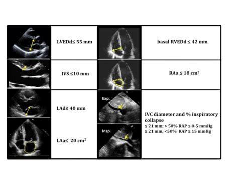
lv dilation echo | dilated left ventricle on echo lv dilation echo Biventricular dilation is associated with a worse prognosis as compared to . Level 1 Level 2 Level 3 Level 4 Level 5 Key affinity condition (unlocked) Increase Trust. (x100) Head to Elmos Square in Tantal.
0 · normal lv end diastolic diameter
1 · lv wall thickness echo
2 · lv dilation echo criteria
3 · left ventricular size on echo
4 · left ventricular diameter chart
5 · left ventricle size chart
6 · dilated left ventricle on echo
7 · dilated cardiomyopathy echo images
Lưu ý: Phương pháp này không thể được sử dụng khi kiểm tra những chiếc túi Vintage được sản xuất trước năm 1980, bởi vì những chiếc túi từ thời đó các mã này sẽ không được tìm thấy và Mã ngày chỉ là mã cho biết sản phẩm được sản xuất ở đâu và khi nào .
Echocardiographic features of DCM are left ventricular (LV) dilation and systolic dysfunction with impaired global contractility and normal LV wall thickness and LV diastolic dysfunction with elevation in LV filling pressure.Biventricular dilation is associated with a worse prognosis as compared to .Two-dimensional transthoracic echocardiography of dilated . Objectives: Identify the etiology and epidemiology of left ventricular hypertrophy and its complications. Outline the appropriate history, physical findings, and evaluation of left ventricular hypertrophy. Summarize the .
normal lv end diastolic diameter
lv wall thickness echo
Assessment of left ventricular systolic function has a central role in the evaluation of cardiac disease. Accurate assessment is essential to guide management and prognosis. Numerous echocardiographic techniques are used in the .Dilated cardiomyopathy (DCM) is defined as left ventricular (LV) dilation and systolic impairment in the absence of coronary artery disease or abnormal loading conditions. Even though robust .
myocardial infarction of the left ventricle (LV) may lead to impaired systolic and diastolic function. The resultant abnormal function exists on a continuum from hypokinesis to akinesis, .
Echocardiography is used to visualize valve anatomy and provide accurate measures of hemodynamic severity, LV hypertrophy, and systolic and diastolic function, and assess for associated conditions, such as aortic .
the dilated LA cavity and LV cavity. The length of postero-basal part of LV compressed by large LA ≥ 30 mm was defined as ‘ giant left atrium’ [45] as shown in Figure 17. Cross sectional . Echocardiographic assessment of left ventricular (LV) diastolic function is an integral part of the routine evaluation of patients presenting with symptoms of dyspnea or heart .The Evaluation of Resynchronization Therapy for Heart Failure and Echocardiography Guided Cardiac Resynchronization Therapy (EchoCRT) trials provided evidence that in patients with .
Echo Case 36: Echocardiography Spot Diagnosis Series - Learn Echo with Labelled findings - Echo Case 36: Echocardiography Spot Diagnosis Series - Learn Echo with Labelled findings .
Echocardiographic features of DCM are left ventricular (LV) dilation and systolic dysfunction with impaired global contractility and normal LV wall thickness and LV diastolic dysfunction with elevation in LV filling pressure. Objectives: Identify the etiology and epidemiology of left ventricular hypertrophy and its complications. Outline the appropriate history, physical findings, and evaluation of left ventricular hypertrophy. Summarize the treatment and management options available for left ventricular hypertrophy.Assessment of left ventricular systolic function has a central role in the evaluation of cardiac disease. Accurate assessment is essential to guide management and prognosis. Numerous echocardiographic techniques are used in the assessment, each .Dilated cardiomyopathy (DCM) is defined as left ventricular (LV) dilation and systolic impairment in the absence of coronary artery disease or abnormal loading conditions. Even though robust data on the epidemiology of DCM are lacking, estimates suggest a disease prevalence of 1:125–250 in adults [1, 2].
myocardial infarction of the left ventricle (LV) may lead to impaired systolic and diastolic function. The resultant abnormal function exists on a continuum from hypokinesis to akinesis, dyskinesis, and aneurysm formation. Of note, the American Society of Echocardiography recommends a wall scoring scale that quantifies the severity of regional wall motion abnormalities (). Echocardiography is used to visualize valve anatomy and provide accurate measures of hemodynamic severity, LV hypertrophy, and systolic and diastolic function, and assess for associated conditions, such as aortic dilatation, mitral valve disease, and elevated pulmonary pressures . 25,34 For patients with AS, repeat echocardiography is .the dilated LA cavity and LV cavity. The length of postero-basal part of LV compressed by large LA ≥ 30 mm was defined as ‘ giant left atrium’ [45] as shown in Figure 17. Cross sectional echocardiography is superior to M-mode echocardiography for measuring left atrial size [46],[47] and it allows the visualization of entire
lv dilation echo criteria
Echocardiographic assessment of left ventricular (LV) diastolic function is an integral part of the routine evaluation of patients presenting with symptoms of dyspnea or heart failure.
The Evaluation of Resynchronization Therapy for Heart Failure and Echocardiography Guided Cardiac Resynchronization Therapy (EchoCRT) trials provided evidence that in patients with mechanical dyssynchrony but narrow QRS duration (< 130 ms), CRT did not improve clinical outcomes or reverse LV remodelling, and instead increased mortality.(53,54)Echo Case 36: Echocardiography Spot Diagnosis Series - Learn Echo with Labelled findings - Echo Case 36: Echocardiography Spot Diagnosis Series - Learn Echo with Labelled findings by Dr. M Usman Javed 3,081 views 1 year ago 1 minute, 20 seconds - Try to spot the findings on the basis of echocardiography, clips shown in the start of the video.
Echocardiographic features of DCM are left ventricular (LV) dilation and systolic dysfunction with impaired global contractility and normal LV wall thickness and LV diastolic dysfunction with elevation in LV filling pressure.
Objectives: Identify the etiology and epidemiology of left ventricular hypertrophy and its complications. Outline the appropriate history, physical findings, and evaluation of left ventricular hypertrophy. Summarize the treatment and management options available for left ventricular hypertrophy.Assessment of left ventricular systolic function has a central role in the evaluation of cardiac disease. Accurate assessment is essential to guide management and prognosis. Numerous echocardiographic techniques are used in the assessment, each .Dilated cardiomyopathy (DCM) is defined as left ventricular (LV) dilation and systolic impairment in the absence of coronary artery disease or abnormal loading conditions. Even though robust data on the epidemiology of DCM are lacking, estimates suggest a disease prevalence of 1:125–250 in adults [1, 2].myocardial infarction of the left ventricle (LV) may lead to impaired systolic and diastolic function. The resultant abnormal function exists on a continuum from hypokinesis to akinesis, dyskinesis, and aneurysm formation. Of note, the American Society of Echocardiography recommends a wall scoring scale that quantifies the severity of regional wall motion abnormalities ().
Echocardiography is used to visualize valve anatomy and provide accurate measures of hemodynamic severity, LV hypertrophy, and systolic and diastolic function, and assess for associated conditions, such as aortic dilatation, mitral valve disease, and elevated pulmonary pressures . 25,34 For patients with AS, repeat echocardiography is .the dilated LA cavity and LV cavity. The length of postero-basal part of LV compressed by large LA ≥ 30 mm was defined as ‘ giant left atrium’ [45] as shown in Figure 17. Cross sectional echocardiography is superior to M-mode echocardiography for measuring left atrial size [46],[47] and it allows the visualization of entire Echocardiographic assessment of left ventricular (LV) diastolic function is an integral part of the routine evaluation of patients presenting with symptoms of dyspnea or heart failure.The Evaluation of Resynchronization Therapy for Heart Failure and Echocardiography Guided Cardiac Resynchronization Therapy (EchoCRT) trials provided evidence that in patients with mechanical dyssynchrony but narrow QRS duration (< 130 ms), CRT did not improve clinical outcomes or reverse LV remodelling, and instead increased mortality.(53,54)

LV Series is a feature-packed, intelligent wall mounted system that includes intelligent eye for energy savings and horizontal auto swing outlet fins for 3-D airflow comfort. With up to 24.5 SEER level, LV Series has been awarded the .
lv dilation echo|dilated left ventricle on echo


